
All about Palmitoyl Tetrapeptide-20 Greyverse in RevivHair REV
All about the remarkable compound Greyverse in remarkable RevivHair REV Advanced with Grey Hair Reverse (product page here):
Product Characteristics
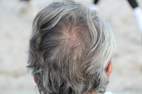
A breakthrough innovation from Lucas Meyer Cosmetics, Greyverse™ is the first anti-grey hair biomimetic peptide. Clinically effective, it helps men and women suffering from greying hair look naturally younger by reversing the hair aging process and increase their self-esteem.
Greyverse™ is the result of a wide screening of several peptides tested on different biological targets involved in the hair greying process. Greyverse™ is a patented Palmitoyl Tetrapeptide (4 amino acids grafted on palmitic acid). The active sequence is derived from the α-MSH structure, known to be the main molecule able to stimulate melanogenesis. As an analogue of α-MSH, Greyverse™ is able to bind to MC1-R (Melanocortin-1 Receptor), a transmembrane receptor located at the surface of melanocytes. The activation of MC1-R stimulates the melanogenesis process, as well as the catalase expression and activity.
Due to its complete mechanism of action, Greyverse™ is able to act simultaneously and efficiently on the two main factors causing hair greying process:
- It stimulates melanogenesis to increase melanin production in the bulb and favor a better transfer leading to hair repigmentation.
- It lowers oxidative stress by increasing the catalase expression and activity, thus reducing the accumulation of hydrogen peroxide to maintain the full pigment color.
Contrary to skin melanocytes, which are able to produce a higher quantity of melanin than the constitutive skin color if they are stimulated (facultative pigmentation), hair melanocytes cannot produce more than they are genetically programmed (constitutive pigmentation only). Thus, Greyverse™ can only help hair recover its natural color, it cannot darken it.
IN VITRO EFFECT OF GREYVERSE™ ON MELANIN SYNTHESIS IN HUMAN MELANOCYTES
- BACKGROUND
Hair pigmentation is due to the presence of a pigment, melanin, secreted by the melanocytes located in the hair bulb and then transferred into the surrounding keratinocytes [1]. The appearance of greying hair is a phenomenon directly linked to a lack of melanin in the hair bulb.
- OBJECTIVE
This study investigated Greyverse™ activity by measuring its effects on the production of melanin in a human melanocyte culture.
- PROTOCOL
Biological materials
The melanocytes used were a cell line of primary normal human epidermal melanocytes from adult tissue (HEMas). HEMas cells were grown in an adherent cell culture realized with CnT-40 CellnTec medium at 37°C, 5% CO2 and 95% humidity.
Tested products
- The Palmitoyl Tetrapeptide-20 (pure peptide contained in Greyverse™) was tested at the following concentration: 10-7M (equivalent to 0.5% Greyverse™).
- The positive control, α-MSH, was tested at a concentration of 10-6 M.
Evaluation of the activity
HEMas cells were seeded in 6-well plates at the concentration of 3.105 cells/mL in a CnT-40 culture medium and incubated at 37°C, 5% CO2. After 24 hours, the medium was removed and the cells were treated with Palmitoyl Tetrapeptide-20 (10-7M) or α-MSH (10-6M) for 72 hours. After 72 hours of treatment, cells were detached for counting, then incubated at 100°C for 10 minutes with NaOH at 1M to extract melanin.

Then, melanin was detected by absorbance at 405 nm. Melanin content was expressed as the percentage of melanin production, compared to cell numbers.
- RESULTS

After 72h of treatment, Greyverse™ significantly increased the melanin synthesis by 19% at the concentration of 10-7 M. The results indicate that Greyverse™ has an activity stronger than the positive control α-MSH.
- CONCLUSION
A biomimetic peptide of α-MSH, Greyverse™ stimulates the melanin synthesis to improve grey hair repigmentation.
EX VIVO EFFECT OF GREYVERSE™ ON PIGMENTATION IN HUMAN HAIR FOLLICLES
- BACKGROUND
Melanosomes are typically divided into four maturation stages (I–IV) determined by their structure and the quantity, quality, and arrangement of the melanin produced.
- Stage I melanosomes have an early matrix organization, are spherical, do not contain tyrosinase activity, and are localized at the periphery of the nucleus.
- Stage II melanosomes are tyrosinase-containing elongated organelles with an organized filamentous matrix. In addition, Mart-1 protein, also known as Melan-A, localized in stage I and/or II, contributes to the melanosome formation.
- Stage III melanosomes, where melanin is uniformly deposited.
- Stage IV melanosomes are melanosomes that are electron-opaque, fully melanized and have a low tyrosinase activity; they are the melanosomes supplied from the dendrites to the neighbouring keratinocytes.
Within melanosomes, at least three key enzymes, tyrosinase, Tyrosinase-related protein 1 (TRP-1), and Tyrosinase-related protein 2 (TRP-2), are absolutely required for the synthesis of different types of melanin. Moreover, the quality of the pigment depends of the balance of the Agouti signaling protein (ASIP, also known as PARD3) and alpha-MSH expression. The binding of ASIP to MC1R precludes alpha-MSH initiated signaling and thus blocks production of cAMP, leading to a down regulation of eumelanogenesis (brown/black pigment) and increasing synthesis of pheomelanin (yellow/red pigment).
- OBJECTIVE
The aim of this study was to determine the effects of Greyverse™ on the pigmentation of isolated human grey hair follicles. To this end, we checked the hair melanosome biogenesis by melanosomal protein immunohistochemistry (Melan-A and TRP-1) and by the nature of the pigment expressed by the immunostaining of ASIP/PARD3.
- PROTOCOL
Tested products
Palmitoyl Tetrapeptide-20 (pure peptide containing in Greyverse™) was tested at the concentrations of 10-9 M and 10-7 M, equivalent to 0.005% and 0.5% Greyverse™, respectively.
Biological materials
On a scalp plasty coming from a 71-year-old Caucasian woman who originally had dark hair, grey hair follicles (from the infundibulum to the bulb) were isolated by micro dissection on day 0 (D0). These hair follicles were placed separately in a 48-well plate and kept in survival for seven days in William’s medium (Sigma-Aldrich, W1878) supplemented with Lglutamine, insulin, hydrocortisone, serum and antibiotics, at 37°C in a 5% CO2 atmosphere. Eleven hair follicles were used per condition (untreated or treated). The Palmitoyl Tetrapeptide-20 was diluted in the culture medium on day 0, day 1, day 4 and day 7 whereas the untreated medium was renewed.
Method
At the end of survival (day 7), the hair shafts were fixed in buffered formalin solution, dehydrated, and paraffin impregnated. 7-µm-thick paraffin sections were mounted on glass slides for immunostainings and stainings.
- For fluorescent immunostainings, the primary antibodies used were anti-PARD3/ASIP (Abcam, ref. ab64646). The sections were then incubated for 30 minutes at room temperature with the Alexa Fluor 488-labeled antibody (Lifetechnologies, ref. A11078). The nuclei were counter-stained using propidium iodide at 0.2µg/mL. The slides were finally mounted using Vectashield® mounting medium (Vector, ref. H-1400).
- For Immunohistochemistry, the hair sections were pre-incubated with hydrogen peroxide (VWR, ref. 23619.264) to inactivate endogenous peroxidase activity. The primary antibodies used were anti-TRP1 (Eurogentec, ref. SIG-38150-1000) and anti-Melan-A (Santa Cruz biotechnologies, ref. sc-20032). Then, the sections were pre-incubated with a biotinylated secondary antibody and incubated with a streptavidin-labeled peroxidase (Vector, Vectastain® Universal ABC kit, ref. PK7200). The staining was revealed by a substrate of peroxidase, VIP (Vector, ref. SK-4600). The nuclei were counter-stained using Mayer hemalun (RAL diagnostics, ref. 320550). The slides were finally mounted using Eukitt mounting medium (VWR, ref. KIND01250).
The microscopic observations were performed using a Leica DMLB or Olympus BX43 microscope. Pictures were digitized with a numeric DP72 Olympus camera with CellD storing software. Different hair parts were analyzed: the infundibulum, upper shaft, bulge, lower shaft and bulb.
- RESULTS

After 7 days of treatments with 10-9 M and 10-7 M Palmitoyl Tetrapeptide-20, the expression of the melanosome protein, Melan-A, was increased especially in the upper sheath and in the bulb area, a site of melanocyte stem cells, attesting an improvement in the melanosome maturation, necessary for an optimal transfer.

Moreover, the expression of the Agouti signaling protein, ASIP/PARD3, was decreased in the lower part of the hair follicle (lower root sheath and bulb). This result indicates that Greyverse™ directs the nature of pigment synthesis: it preferentially increases eumelanin synthesis over pheomelanin synthesis, allowing the natural dark hair color of the donor to be recovered.

The key enzyme of melanin synthesis, tyrosinase-related proteins 1 (TRP1), was induced in the upper root sheaths, attesting the greater capacity to produce pigments.
- Conclusion
Greyverse™ promotes hair pigmentation by increasing melanosome transfer and melanin synthesis, preferably eumelanin.
IN VITRO EFFECT OF GREYVERSE™ ON MELANOSOME TRANSFER IN A CO-CULTURE OF KERATINOCYTES AND MELANOCYTES
- BACKGROUND
Hair pigmentation can be influenced by the concentration of melanin as well as by the number, size and distribution of melanosomes around the nucleus of keratinocytes. Melanin is synthesized, stored and transported within a specialized organelle called a melanosome in melanocyte. The transfer of melanin from melanocytes to keratinocytes (melanosome transfer + phagocytosis) is a critical step in skin pigmentation, one that depends on various parameters such as melanocyte dendricity.
- OBJECTIVE
n this study, the potential pro-pigmenting effect of Greyverse™ was evaluated in a co-culture of normal human epidermal keratinocytes (NHEK) and normal epidermal melanocytes (NHEM). In brief, the effect of Greyverse™on melanosome transfer was evaluated using flow cytometry to quantify a melanosome specific marker (Pmel17) inside keratinocytes identified by the expression of the cell surface marker CD49f.
- PROTOCOL
Tested products
- Palmitoyl tetrapeptide-20 (pure peptide contained in Greyverse™) was tested at the concentration of 10-9M (equivalent to 0.005% Greyverse™)
- Positive reference: stimulation mix containing pro-pigmenting factors prepared in accordance with the internal protocol of Bioalternatives (Gençay, France).
Biological materials
The human keratinocytes and human melanocytes were seeded (10:1 ratio) in 24-well plates in a co-culture medium and incubated for 48 hours, with a medium renewal after 24 hours. The medium was then removed and replaced by a co-culture medium containing or not containing the active compound or the positive reference. The cells were then incubated for 48 hours. Half of the medium was then discarded, the treatments were renewed and the cells were incubated for 72 hours. All experimental conditions were performed in n=4.
Labelling and cytometry analysis
At the end of the incubation period, the medium was removed and cells were rinsed with a PBS solution. Cells were then detached using a trypsin treatment and transferred to tubes. After several washes in PBS/BSA 0.2% cells were fixed, permeabilized and labeled with the antibodies below:
- Anti-CD49f-FITC: CD49f corresponding to integrin α6 specific to keratinocytes
- Anti-Pmel17 (specific to melanocytes and melanosomes). Anti-Pmel17 labeling was revealed using a conjugated allophycocyanin (GAM-APC) secondary antibody
- Or both.
Acquisitions were performed on 10,000 cells for each replicate, after selection of a homogeneous population analyzed for size (FSC) and granularity (SSC) parameters by flow cytometry using a BD FACSVerse™ flow cytometer. Data analysis was carried out using FlowJo vX software. The melanosome transfer evaluation was performed by quantifying the double-labeled keratinocyte population for CD49f and Pmel17 and calculating the percentage of variation compared to the untreated control.

Statistical Analysis
The inter-group comparisons were performed using an Unpaired Student’s t-Test: *p<0.05, **p<0.01 and ***p<0.001.

After 72 hours of treatment, Greyverse™ significantly increased the melanosome transfer between melanocyte and keratinocyte cells by 50%.
- CONCLUSION
Greyverse™ stimulates melanosome transfer, thus increasing melanin content in the hair shaft and leading to natural hair pigmentation recovery.
EX VIVO EFFECT OF GREYVERSE™ON MACROSCOPIC PIGMENTATION IN ISOLATED HUMAN HAIR FOLLICLES
- OBJECTIVE
To complete the previous data, the aim of this study was to evaluate the activity of Greyverse™ on the macroscopic hair pigmentation of isolated hair follicles maintained in survival.
- PROTOCOL
Tested products
Palmitoyl Tetrapeptide-20 (pure peptide contained in Greyverse™) was tested at the concentrations of 10-9M and 10-7M, equivalent to 0.005% and 0.5% Greyverse™, respectively.
Biological materials
On a scalp plasty coming from a 76-year-old woman, 74 inferior hair follicles (lower root sheath and bulb) were isolated by microdissection. These hair follicles were placed in 48-well plates and kept in survival for 8 days at 37°C in a 5% CO2 atmosphere, in a Philpott medium composed of William’s medium (ref. W1878, SigmaAldrich) supplemented with L-Glutamine, insulin-transferrin-selenium, hydrocortisone, penicillin/streptomycin antibiotics and serum (300 μl per well).
Method
The entire culture medium was renewed (300 μl per well) on day 0, day 1, day 4 and day 6 and the products to be tested were added to it. The untreated batch did not receive any treatment other than the renewal of the medium.
Hair follicle pigmentation analyses
Microscopic observation of grey hair follicle pigmentation:
The method consisted in mounting selected hairs on clear glass slides for microscopic observation (hairs were then fixed)
- Hair pigmentation on mounted hairs
Selected hair follicles were fixed and mounted on clear glass slides using a Clarine-Eukitt mounting medium to visualize the hair pigmentation by microscopic observation. The microscopic observations were performed using a Leica DMLB or Olympus BX43 microscope. Pictures were digitized with a numeric DP72 Olympus camera with CellD storing software.

Picture analysis
All image processing was performed with Fiji software [2]. The first part of the processing allowed the hair follicle to be extracted from the rest of the image and its surface measured (in mm²). The “subtract background” function was used to remove smooth continuous backgrounds from the image. Then, the image intensities were enhanced using the Enhanced Local Contrast (CLAHE) plugin. This filter allows the local contrast in an image to be equalized (it reduces inhomogeneity in terms of intensity), while preserving the selected regions of interest and limiting the amplification of noise. This method helped increase hair detection. Finally, an automatic threshold level was set to create the corresponding binary image of the hair, and calculate area statistics (hair surface in mm²).
In the second part of the processing, an automatic thresholding was used on each raw image in order to select pixels corresponding to the pigmented area only. Using binary images of the selected hair (first part of the pipe) as an inclusive mask, the surface of these pigments (in mm²) was computed in the hair area only.

Statistical Analysis
The inter-group comparisons were performed by an Unpaired Student’s t-Test: *p<0.05, **p<0.01 and ***p<0.001.
- RESULTS
The micro-dissected hair follicles were put in survival and photographed alive (20 hairs per batch) on day 0 (before treatment) and day 8 (after treatment). The evolution of the hair bulb pigmentation was estimated by image quantification on day 8 compared to day 0, for each isolated hair follicle. The results were expressed as a percentage of variation of area hairs exhibiting an increase in hair pigmentation.
According to the experimental conditions, after 8 days of survival and compared to day 0, the Palmitoyl Tetrapeptide-20 Fgrey increased hair pigmentation by 52% and 66% at concentrations of 10-9 M and 10-7M, respectively.


The color of the treated hair follicles had increased, attesting a higher melanin content.
- CONCLUSION
Greyverse™ visibly darkens hair by increasing the melanin content in the hair shaft.
IN TUBO EFFECT OF GREYVERSE™ ON CATALASE ENZYMATIC ACTIVITY
- BACKGROUND
Recently, Shi and al. [3] have shown that a deficiency of catalase, an enzyme that allows the transformation of hydrogen peroxide into oxygen and water (2 H2O2 → 2 H2O + O2) in human hair follicles, largely contributes to the greying of hair. Moreover, it was shown that it induces a change in the conformation of the tyrosinase structure synthesized by hair follicle melanocytes, especially by the oxidation of the methionine residues of this key enzyme in melanin production. This structural modification of the tyrosinase leads to a loss of its enzymatic activity and stops melanin synthesis in the melanocytes of the hair follicle.
- OBJECTIVE
The aim of this study was to determine the direct effect of Greyverse™ on catalase activity by measuring the quantity of H202 not degraded by the enzyme.
- PROTOCOL
Tested products
Palmitoyl Tetrapeptide-20 was tested at the concentration of 10-5 M.
Method
The catalase activity was evaluated using the Amplex® Red Catalase Assay Kit purchased by Molecular Probes (Invitrogen, ref: A22180) in compliance with the instructions of the manufacturer. In the assay, catalase first reacts with H2O2 to produce water (H20) and oxygen (O2). Then, the Amplex Red reagent reacts with a 1:1 stoichiometry with any unreacted H2O2 in the presence of horseradish (HRP) to produce the highly fluorescent oxidation product, resorufin. A correlation with catalase activity is then calculated.

A 10-5 M Palmitoyl Tetrapeptide-20 solution was incubated with 6.25 mU catalase in a 96-well plate for 10 minutes at room temperature. Then, an extemporaneously prepared solution of H2O2 (20 µM final concentration) was added and the mixture was incubated for 30 minutes at room temperature in the dark. A mixture of HRP (0.2U/mL final concentration) and Amplex Red fluorogenic probe (50 µM final concentration) was then added and the plates were incubated in the dark at 37°C for 60 minutes.
Evaluation of the activity
The generated fluorescence of formed resorufin was immediately recorded (λex = 544nm, λem = 590 nm) using a Polar Star Omega (BMG, Ortenberg, Germany) reader.
Statistical Analysis
The inter-group comparisons were performed by an Unpaired Student’s t-Test: #p<0.1, *p<0.05, **p<0.01 and ***p<0.001.
- RESULTS

The Palmitoyl Tetrapeptide-20 is able to significantly increase, in tubo, catalase activity by 7.5%.
- CONCLUSION
Greyverse™ improves catalase activity to reduce the greying hair process due to oxidative stress caused by excess H202.
IN VITRO EFFECT OF GREYVERSE™ ON THE INTRACELLULAR H2O2 LEVEL IN HAIR FOLLICLE DERMAL PAPILLA FIBROBLASTS
- BACKGROUND
Recently, Shi and al. have shown that a deficiency of catalase, an enzyme that allows the transformation of hydrogen peroxide into oxygen and water (2 H2O2 → 2 H2O + O2) in human hair follicles, largely contributes to the greying of hair.
- OBJECTIVE
The aim of this study was to determine the effect of Greyverse™ on the intracellular H2O2 level due to its action on the increased catalase activity.
- PROTOCOL
Tested products
- Palmitoyl Tetrapeptide-20 was tested at the concentration of 10-5 M.
- The 2’,7’-Dichlorofluorescein diacetate (DCFH-DA) was used at a final concentration of 10 µM.
Biological materials
HFDP cells are fibroblasts isolated from human hair dermal papilla and maintained in a specific follicle dermal papilla cell medium (Promocell, Ref: C-26501) containing fetal serum (0.04 mL/mL), bovine pituitary extract (0.004 mL/mL), basic fibroblast growth factors (recombinant human, 1 ng/mL), insulin (recombinant human, 5µg/mL) and antibiotics (penicillin/streptomycin/Eurobio, CABPES01-OU) at 37 °C under 5% CO2 and 95% humidity.
Method
The intracellular levels of H2O2 were evaluated by using a fluorescent dichlorofluorescein assay adapted to flow cytometry which allowed the detection of picomole levels of hydroperoxides [4]. Using the fluorogenic probe and the 2’,7’-Dichlorofluorescein diacetate (DCFH-DA) (Sigma, Ref: D6883), we assessed the levels of intracellular hydrogen peroxide within the untreated control and the treated cells. HFDPC cells were seeded at 2.106 cells/well in 12-well plates with 500 µl complete medium and incubated for 24 hours at 37°C. Palmitoyl Tetrapeptide-20 (10-5 M) was then added and incubated for 18 hours at 37°C. At the end of incubation, DCFH-DA probe (10 µM final concentration) was added for an additional 15 minutes.
Evaluation of the activity
Cells were then trypsinized, washed and immediately scanned on an FACS (FC500 flow cytometer, BeckmanCoulter) with excitation and emission settings of 485 nm and 529nm, respectively. The emitted fluorescence is assumed to be proportional to the concentration of hydrogen peroxide in the cells [5]. The results are expressed as a percentage of decrease in the H2O2 production by HFDPC in comparison with the untreated control cells.
Statistical Analysis
The inter-group comparisons were performed by an Unpaired Student’s t-Test: *p<0.05, **p<0.01 and ***p<0.001.
- RESULTS

Greyverse™ is able to decrease the internal H2O2 production in an HFDPC culture by 30%.
- CONCLUSION
Greyverse™ decreases H2O2 accumulation thus decreasing oxidative stress involved in the greying hair process.
EX VIVO EFFECT OF GREYVERSE™ ON OXIDATIVE ENZYME EXPRESSION
- BACKGROUND
Oxidative stress is one of the main causes of the greying process. The excess of free radicals degrades melanin and lowers the capacity of the melanocytes to synthetize melanin. Shi and al. have shown that a deficiency of catalase, the enzyme that allows the transformation of hydrogen peroxide into oxygen and water (2 H2O2 → 2 H2O + O2) in human hair follicles, largely contributes to the greying of hair [3]. Moreover, TRP-2 expression was shown to be uncoupled to that of tyrosinase and TRP-1 in hair bulb melanocytes, whatever the hair colour and ethnic origin [6], suggesting a specific role of this enzyme apart from pigmentation through melanin synthesis. Michard et al. investigated the TRP-2 effects on oxidative stress-mediated toxicity [7]. They demonstrated that TRP-2 over-expression reduces H2O2-induced DNA damage, with a significant improvement of respiration and survival rate without any impact on cell proliferation. Additionally, they demonstrated that TRP-2 silencing partially restores sensitivity to oxidative stress. The importance of TRP-2 as a protector for hair follicle and hair colour preservation is further confirmed by the fact that TRP-2 expression is preserved in eyelash follicle melanocytes [8]: it would explain eyelash protection against greying. Altogether, we can seriously presume that catalase and TRP-2 contribute to protecting hair follicle melanocytes from oxidation, and that their specific lack leads to hair follicle susceptibility to oxidation [9] and largely contributes to the greying of hair.
- OBJECTIVE
The aim of this study was to determine ex vivo the effects of Greyverse™ on oxidative enzymes (catalase and TRP-2) implicated in the greying hair process.
- PROTOCOL
Tested products
Palmitoyl Tetrapeptide-20 (pure peptide contained in Greyverse™) was tested at concentrations of 10-9 M and 10-7M, equivalent to 0.005% and 0.5% Greyverse™, respectively.
Biological materials
On a scalp plasty coming from a 71-year-old Caucasian woman, grey hair follicles (from the infundibulum to the bulb) were isolated by microdissection on day 0. These hair follicles were placed separately in a 48-well plate and kept in survival for seven days in William’s medium (Sigma-Aldrich, W1878) supplemented with L-glutamine, insulin, hydrocortisone, serum and antibiotics, at 37°C in a 5% CO2 atmosphere. Eleven hair follicles were used per condition (untreated or treated). Palmitoyl Tetrapeptide-20 was diluted at 10-9 M and 10-7 M in the culture medium on day 0, day 1, day 4 and day 7 whereas the medium of the untreated hair follicles was renewed.
Method
At the end of survival (day 7), hair follicles were fixed in a buffered formalin solution, dehydrated, and paraffin impregnated. 7-µm-thick paraffin sections were mounted on glass slides for immunostainings and stainings.
- For fluorescent immunostainings, the primary antibody used was i-PARD3/ASIP (Abcam, ref. ab64646). The sections were then incubated for 30 minutes at room temperature with the Alexa Fluor 488-labeled antibody (Lifetechnologies, ref. A11078). The nuclei were counter-stained using propidium iodide at 0.2µg/mL. The slides were finally mounted using Vectashield® mounting medium (Vector, ref. H-1400).
- For Immunohistochemistry, the hair sections were pre-incubated with hydrogen peroxide (VWR, ref. 23619.264) to inactivate endogenous peroxidase activity. The primary antibodies used were anti-TRP1 (Eurogentec, ref. SIG-38150-1000) and anti-Melan-A (Santa Cruz biotechnologies, ref. sc-20032). Then, the sections were pre-incubated with a biotinylated secondary antibody and incubated with a streptavidin-labeled peroxidase (Vector, Vectastain® Universal ABC kit, ref. PK7200). The staining was revealed by a substrate of peroxidase, VIP (Vector, ref. SK-4600). The nuclei were counter-stained using Mayer’s hemalum (RAL diagnostics, ref. 320550). The slides were finally mounted using Eukitt mounting medium (VWR, ref. KIND01250).
The microscopic observations were performed using a Leica DMLB or Olympus BX43 microscope. Pictures were digitized with a numeric DP72 Olympus camera with CellD storing software. Different hair parts were analyzed: infundibulum, upper shaft, bulge, lower shaft and bulb.
- RESULTS
After 7 days of treatments with 10-9 M and 10-7 M Palmitoyl Tetrapeptide-20, an antioxidant activity was observed by immunostainings on micro-dissected human hair follicles.

The tyrosinase-related proteins TRP-2 was induced in the upper root sheaths.

The antioxidant enzyme catalase, known to block the deleterious whitening effect of hydrogen peroxide in hair, was increased in the lower root sheaths.
- CONCLUSION
Greyverse™ decreases the enzymes involved in oxidative stress thus reducing the greying hair process.
REF: Knowde.com